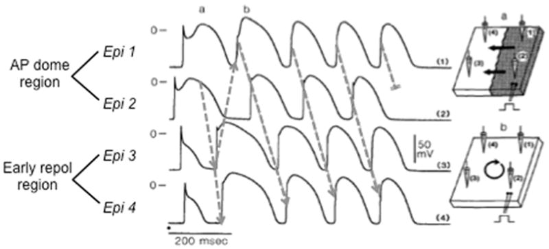
Figure 5.

Phase 2 reentry during simulated ischemia in canine epicardium. Traces show AP recordings from four sites (Epi 1–4) in a canine epicardial sheet exposed to simulated ischemia ([K+]o=6 mmol/l, hypoxia, pH=6.8). Sites 1 and 2 exhibit normal APs with accentuated AP domes, whereas sites 3 and 4 show early repolarization. Arrows show re-excitation of site 3 by the AP dome at site 2, inducing phase 2 reentry that self-terminates after 4 beats. Adapted from Lukas & Antzelevitch 42.
