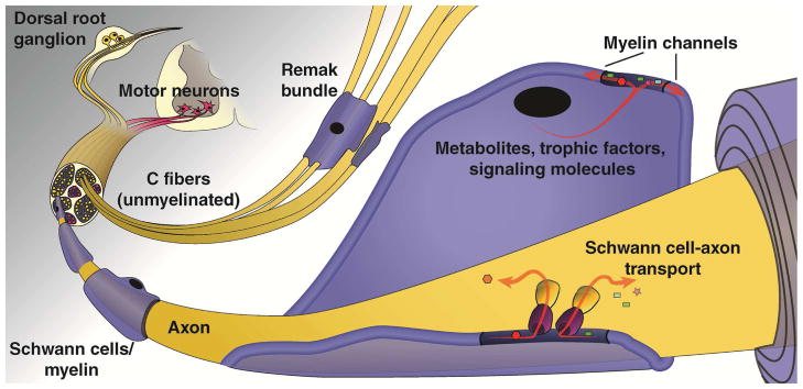
Figure 1. The peripheral nervous system.
The peripheral nervous system (PNS) is comprised of both neurons and Schwann cells (SCs), and the structure, location, and interaction of these components have important implications for PNS function. Efferent axons of motor neurons, whose cell bodies are located in the ventral horn of the spinal cord, carry signals from the central nervous system (CNS) to muscles and glands, whereas afferent axons of sensory neurons, whose cell bodies are located in the dorsal root ganglia, relay information from peripheral sensory receptors to the CNS. Thin and unmyelinated sensory axons, also known as C-fibers or small fibers, are associated with non-myelinating SCs and are grouped as Remak bundles and represent a large portion of the PNS neurons. Myelinated sensory axons, on the other hand, are surrounded by myelin sheaths made by SCs that form distinct nodal domains important for saltatory conduction and a tubular network of myelinic channels that connect the SC cytoplasm with the periaxonal space to provide a source of energy to the axonal compartment

