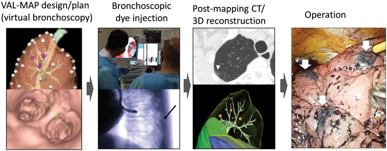
Figure 1:
Steps of VAL-MAP. The lung ‘map’ was designed using radiology workstations and virtual bronchoscopy. Bronchoscopic dye injection was conducted within 3 days before surgery under fluoroscopic guidance to confirm the location of the metal-tip injection catheter (black arrow). After mapping, CT scan was taken within a few hours–days after VAL-MAP to visualize actual locations of markings (arrowhead). Using a radiology workstation, 3D images were further reconstructed, reflecting actual locations of markings. The operation was conducted using the 3D image for guidance. The white arrows indicate dye marks. VAL-MAP: virtual-assisted lung mapping.

