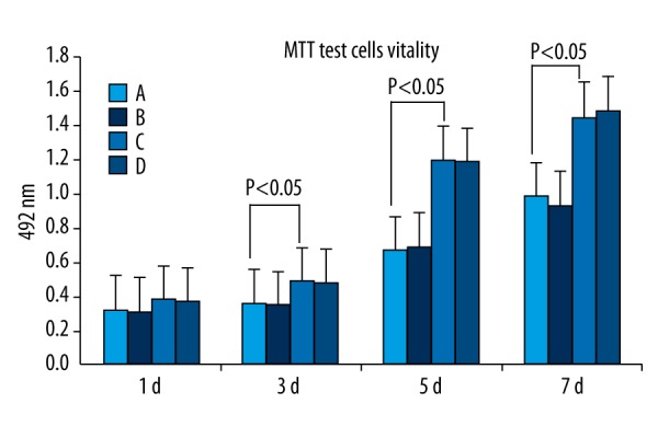
Figure 3.

MTT assay of untransfected PMSCs (A) and untransfected BMSCs (B) and GDNF-transfected PMSCs (C) and GDNF-transfected BMSCs (D). After 3–5 d of transfection, PMSCs and BMSCs exhibited logarithmic growth and the growth rate slowed at 6–7 d. The cell viability of all 4 groups was not significantly different (P>0.05) at 24 h after transfection. At 3 d after transfection, the cell viability of GDNF-transfected-PMSCs and GDNF-transfected-BMSCs gradually became significantly higher than that of PMSCs and BMSCs (P<0.05). The cell viability of GDNF-transfected-PMSCs and GDNF-transfected-BMSCs was not significantly different (P > 0.05).
