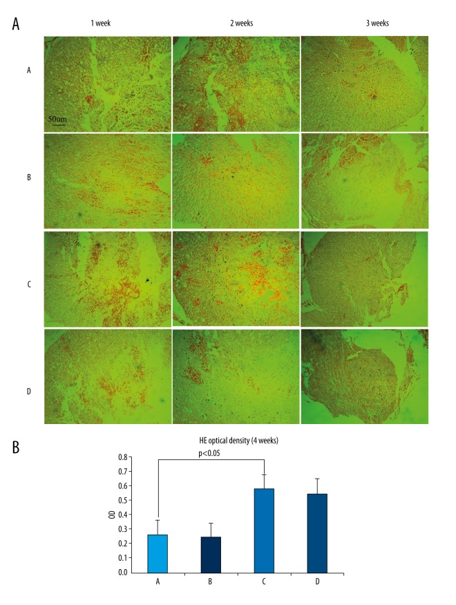
Figure 6.
(A, B) HE staining observed under inverted phase-contrast microscopy (×100). One week after surgery, inflammatory cells, cavities, and nerve fiber necrosis can be observed in the SCI. Nerve cell proliferation can be also observed in GDNF-transfected-PMSCs (C) and GDNF-transfected-BMSCs (D). After 2 weeks and in all 4 groups, the SCI was filled with dense fibrous connective tissue. Astrocyte proliferation and accumulation in the glial scar is visible. There were obviously more proliferating cells in GDNF-transfected-PMSCs (C) and GDNF-transfected-BMSCs (D) than in PMSCs (A) and BMSCs (B). After 4 weeks, the scar tissue can still be observed in each group but with reduced cysts and necrosis. The tissue is denser, while there are significantly increased nerve cells as compared to that at 2 weeks. There was significantly increased nerve cell proliferation in GDNF-transfected-PMSCs (C) and GDNF-transfected-BMSCs (D) but it was much lower in PMSCs (A) and BMSCs (B). Overall, GDNF-transfected-PMSCs (C) and GDNF-transfected-BMSCs (D) did not differ greatly, but were significantly improved as compared to PMSCs (A) and BMSCs (B).

