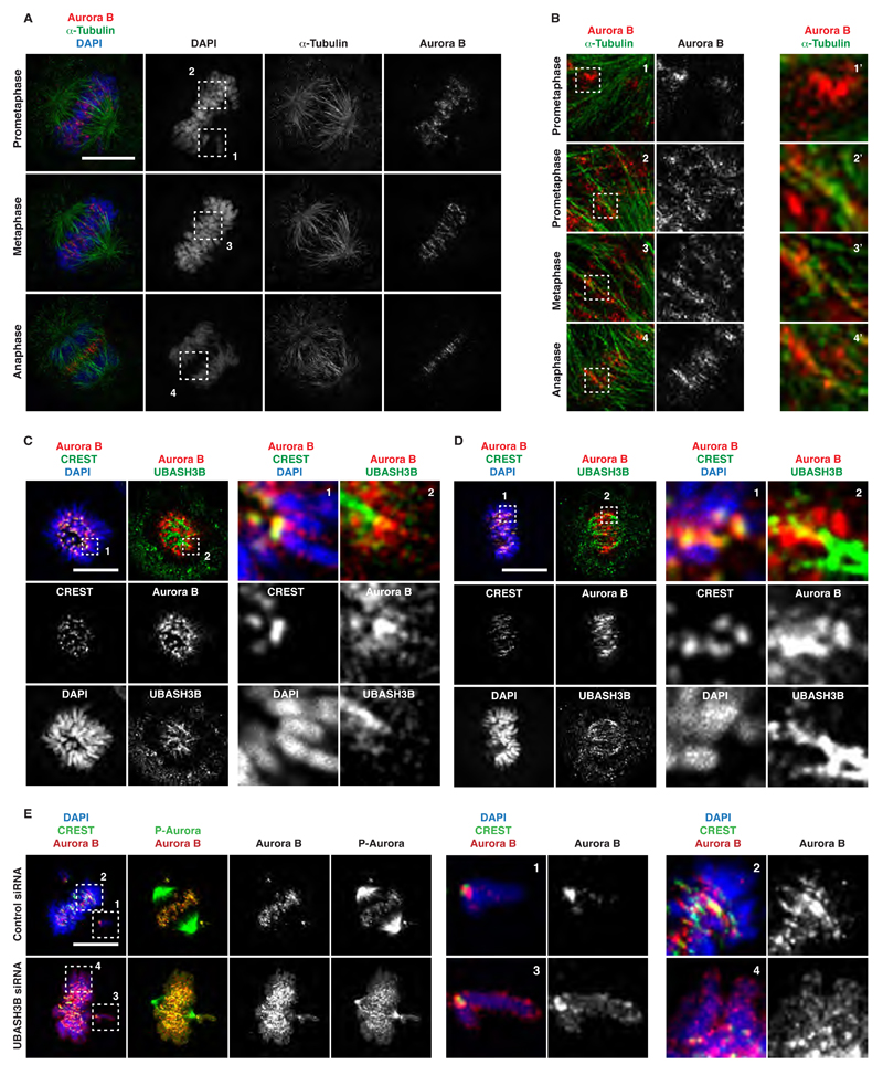
Figure 6. UBASH3B mediates the microtubule localization of Aurora B upon chromosome alignment.
(A and B) HeLa cells were synchronized in different mitotic stages (prometaphase, metaphase, anaphase) by a double thymidine block and release and analysed by super-resolution microscopy. The framed and numbered regions in (A) are magnified and depicted in the corresponding panels in (B). These regions are further magnified in the corresponding right panels (indicated by the corresponding numbers with ’). Hereafter, scale bar is 5 μm. (C and D) MDA-MB-231 cells were synchronized by a double thymidine block and release in different mitotic stages (early in C and late prometaphase in D) and analysed by immunofluorescence microscopy (IF). The magnified framed and numbered regions are shown in the corresponding right panels. (E) MDA-MB-231 cells were treated with control and UBASH3B siRNAs, synchronized as in (C, D) and analysed by IF. The magnified framed and numbered regions are shown in the corresponding right panels.

