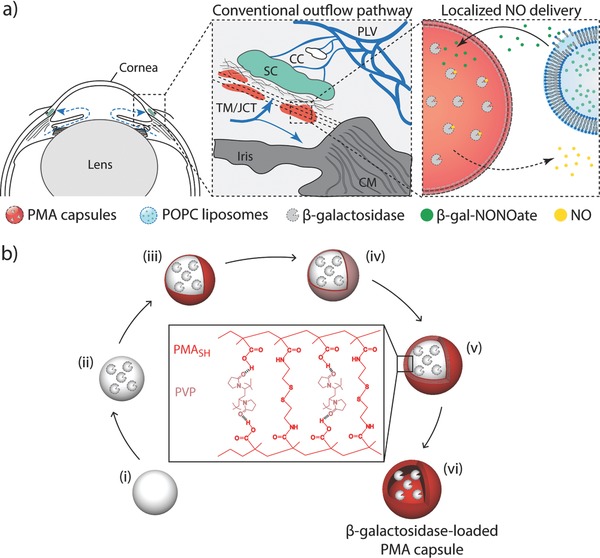
Scheme 1.

a) Schematic illustration of localized delivery of nitric oxide (NO) to the conventional outflow pathway via enzyme biocatalysis. Left: Anterior segment of eye showing the direction of aqueous humor flow in blue. Center: Enlargement of the iridocorneal angle (boxed region in left panel) showing the conventional outflow pathway. Right: Schematic of localized NO delivery within the trabecular meshwork (TM) near Schlemm's canal (SC). β‐Galactosidase is encapsulated in poly(methacrylic acid) (PMA) capsules and enmeshed within the TM. Liposomes containing NO donors (β‐gal‐NONOate) are delivered to the outflow pathway. Upon liposome degradation, NO donors are slowly released at the outflow resistance sites and enzymatic activity of β‐galactosidase results in local delivery of active therapeutic NO at the outflow resistance sites, achieving a targeted on‐site NO delivery to the conventional outflow pathway. CC: collector channels, CM: ciliary muscle, JCT: juxtacanalicular connective tissue, and PLV: perilimbal vessels. b) Schematic illustration of assembly of β‐galactosidase‐loaded PMA capsules via layer‐by‐layer technique. i) Aminated silica particle template is coated with ii) β‐galactosidase, followed by sequential deposition of iii) thiol‐functionalized PMA (PMASH) and iv) poly(N‐vinylpyrrolidone) (PVP) via hydrogen bonding. v) Once four bilayers of PMASH/PVP are deposited, the thiol groups of PMASH are oxidized into bridging disulfide linkages. vi) Removal of the sacrificial particle template results in (bio)degradable disulfide‐cross‐linked β‐galactosidase‐loaded PMA capsule.
