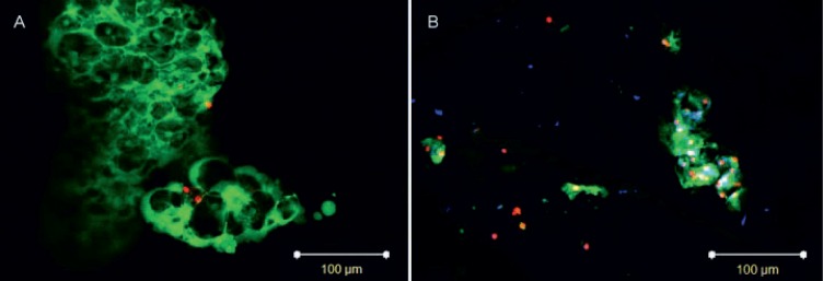
Fig. 2.
Notochordal cells (NCs) (A) prior to injection and (B) in nucleus pulposus tissue after 42 days of culture. Cytoplasm of NCs is stained in green (cell tracker carboxyfluorescein diacetate succinimidyl ester (CFDA-SE)), cytoplasm of all cells in blue (calcein blue-AM; only in B), and nuclei of dead cells in red (propidium iodide). Scale bars = 100 μm. Representative of n = 6.

