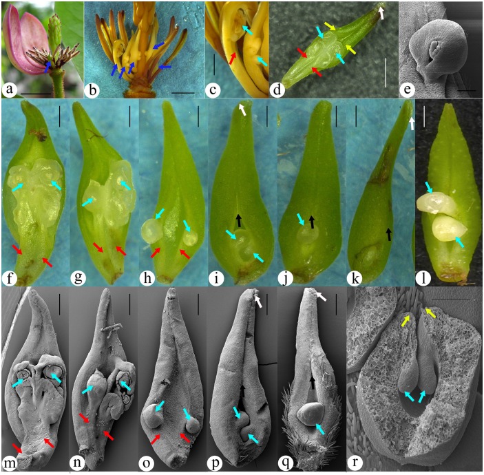
Fig 1. Configuration of atypical female units that are transitional to the typical one.
A-K are stereomicrography, L-S are SEM. (A) A post-anthetic flower with an atypical female unit (arrow) situated between the male and female sections. The tepals are removed to show inner flower parts. Bar = 10 mm. (B) A flower with several atypical female units (arrows) between the male and female sections. Bar = 10 mm. (C) Detailed view of the atypical female units shown in Fig. 1B. Note the exposed ovules and that, in at least one female unit, the ovuliferous branch (placenta) is obviously separated (arrow) from the subtending foliar part. Bar = 1 mm. (D) Adaxial view of an atypical female unit comprising a subtending foliar part and a placenta in its axil. Papillae (blue arrow) and enrolling margins (white arrow) are seen on the distal of the foliar part. The placenta comprises two slightly fused branches (red arrows), each of which terminates in an ovule (yellow arrow). Bar = 1 mm. (E) One of the ovules in Fig. 1m that is attached to the placenta (red arrow) isolated from the subtending foliar part (black arrow). Bar = 0.5 mm. (F-Q) A serial pairs of LM and SEM images showing female units transitional from atypical to typical configuration. The spatial relationship between the ovules and subtending foliar parts changing from isolated gradually into increasingly fused, and the presence of ovuliferous branch is increasingly hard to see. Figs. 1f-j and 1m-q are from a single flower. Bar = 0.5 mm. (F, M) Anatypical female unit with a configuration similar to that in Fig. 1D. Note the barely fused branches (red arrows, placenta) terminating in ovules (yellow arrows). One of the ovule is shown in detail in Fig. 1e. (G, N) Ovules (yellow arrows) on the tips of branches (placenta, red arrows) subtended by a foliar part. (H, O) Two ovules (yellow arrows) appearing borne on the margins of the foliar part due to the fusion between the two branches (of placenta, red arrows) and foliar part margins. The foliar part has its margins (white arrow) enrolled in the distal portion. (I, P) Further enrolling of the foliar part giving rise to an obvious ventral suture (white arrow). Note the ovules (yellow arrows) are more enclosed than in Fig 2g and 2n. (J) Almost completely closed female unit with obvious ventral suture (white arrow) and only one ovule (yellow arrow) visible. (K) A completely closed female unit. Its ovule (yellow arrow) is fully enclosed and visible only when the female unit is cut at the bottom. (L) A closed female unit that fails to enclose its ovules. (Q) Almost completely closed female unit with obvious ventral suture (white arrow) and only one ovule (yellow arrow) visible. (R) A cross-cut typical female unit showing the ovules (yellow arrows) fused to the margins (white arrows) of the foliar part. Only this image was slightly horizontally squashed to fit into the space available. Bar = 0.2 mm.

