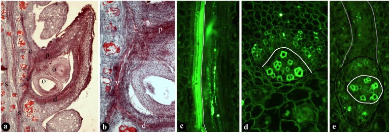
Fig 2. Anatomy of typical fruits showing vascular bundles in the fruit wall and placenta.
(A) Longitudinal radial section of a fruit showing dorsal (d) and ventral (v) bundles, and placenta bundle (p) supplying the ovules (o). Bar = 1 mm. (B) Detailed view enlarged from Fig. 2a, showing dorsal bundle (d), ventral bundle (v), placenta bundle (p) supplying the ovules (arrows). Bar = 0.1 mm. (C) Longitudinal section of a collateral dorsal bundle in the fruit wall, showing adaxial xylem (to the left of white line) and abaxial phloem (to the right of white line). Bar = 50 μm. (D) Cross view of a collateral stellar bundle (black line) with adaxial xylem (below white line) and abaxial phloem (above white line). Bar = 20 μm. (E) Cross view of an amphicribral placenta bundle (black line) with xylem (within in the white line) surrounded by phloem (between the white and black lines). The bundles is extended (gray line) above to an ovule. Bar = 20 μm.

