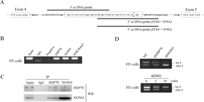
Fig 3. Effects of proteins associated with the cryptic exon area in Int4 of GLA.
(A) Schematic illustration of GLA with positions of the biotin-labelled DNA probes for pull-down assays. (B) ChIP analysis on the cryptic exon area in Int4 of GLA was performed using antibodies against HSP70, NONO, and H3K36me3 with IgG as a control. (C) Co-immunoprecipitation results using anti- HSP70 or anti-NONO antibody for immunoprecipitation and analyzing by western blotting. Nonimmune IgG was used as negative control. (D) Fabry disease cells were infected with lentiviruses expressing shRNAs targeting HSP70 or NONO, or treated with 4β-hydroxywithanolide E (4HWE). Messenger RNA was extracted after 48 hours infection or 24 hours 4HWE treatment followed by RT-PCR analysis. WB, western blot; NC: negative control.

