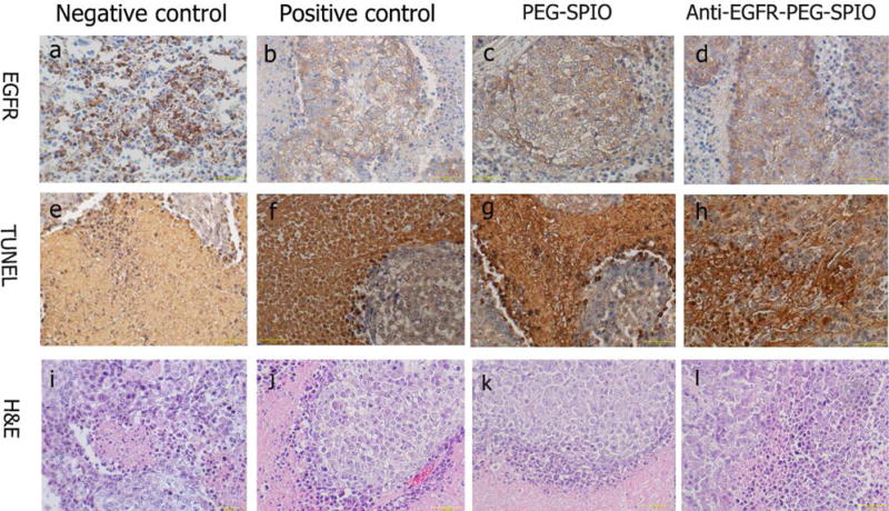
Fig. 8.

The expression of EGFR in tumor tissue after MRgFUS ablation by immunohistochemistry. The nucleus appeared brown (a) in EGFR-positive cells, blue in negative cells (b and c and d). TUNEL staining images showed that the percentage of apoptotic cells in (e) were much lower than those in (f and j and h). (i~l) H&E staining of tumor tissue in negative control (low power), positive control, PEGylated SPIO and anti-EGFR-PEG-SPIO group.
