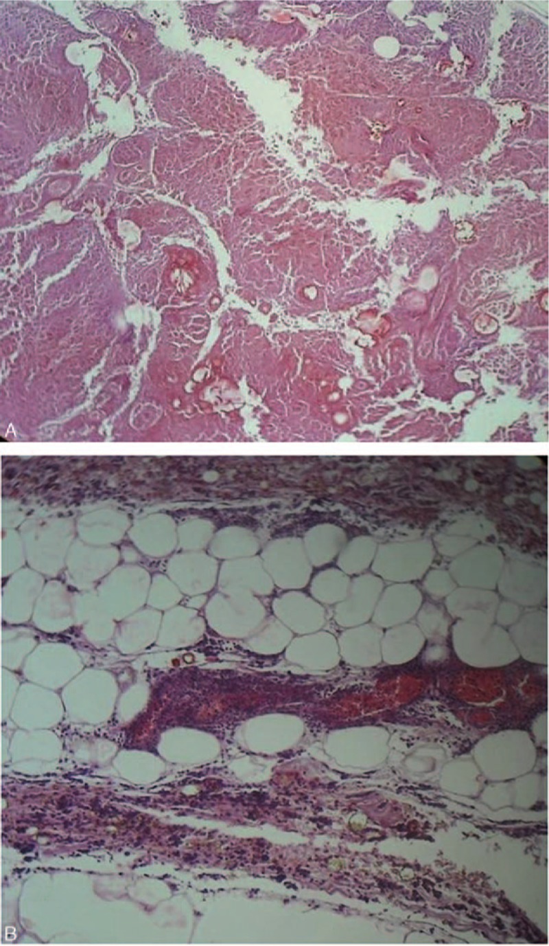
Figure 4.

Microscopic view of the pancreas showing (A) the pancreatic tissue totally necrosis and mostly overwhelmed with extravasated blood (hematoxylin–eosin staining, original magnification, ×40), and (B) the extension into the peripancreatic adipose tissue with numerous polynucleated white blood cells (hematoxylin–eosin staining, original magnification, ×100), suggestive of severe hemorrhagic acute pancreatitis.
