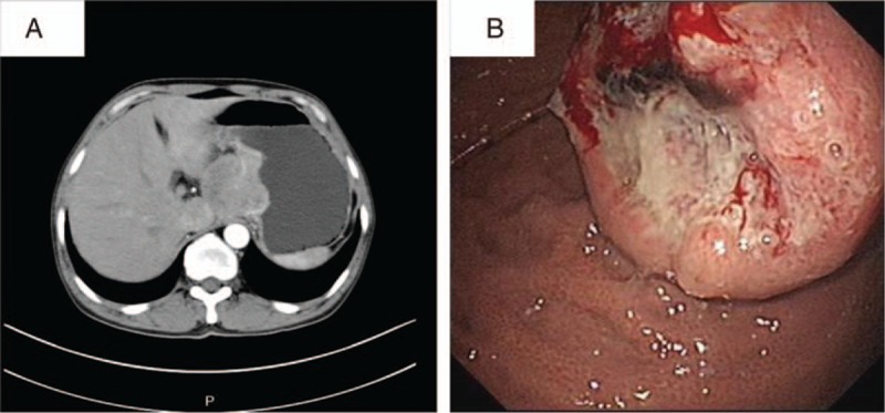
Figure 2.

(A) Abdominal enhanced computed tomography (CT) scan images of a 50-year-old male patient with gastric SCC treated in our hospital showed a mass of 5.0 × 4.8 cm in size in the fundus of stomach, protruding to hepatogastric space. (B) Electronic gastroscopy revealed a gray-white and fragile tumor-like bulge of 4.0 × 4.0 cm in size with irregular surrounding mucosa. SCC = squamous cell carcinoma.
