. 2017 Mar 21;6:e22512. doi: 10.7554/eLife.22512

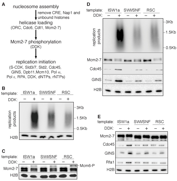
Figure 4. SWI/SNF and RSC templates show reduced CMG formation.

(A) Outline of fully-reconstituted nucleosomal DNA replication initiation assay. The proteins added at each step are indicated. (B) Comparison of reconstituted nucleosomal DNA replication using ISW1a, SWI/SNF and RSC templates in the presence or absence of DDK. Analysis of replication products and H2B as in Figure 3B. (C) Comparison of Mcm2-7 phosphorylation by DDK on ISW1a, SWI/SNF and RSC templates. Phosphorylation of Mcm6 is indicated by reduced electrophoretic mobility and was analyzed by immunoblot (top). Template associated H2B is shown (lower). (D) Comparison of replication of ISW1a, RSC and SWI/SNF templates. Reactions were performed with or without DDK and replication products of the reconstituted replication reactions were analyzed as in Figure 3B (top). Template association of Mcm2-7, Cdc45, GINS and H2B was measured after a high-salt wash at the end of reconstituted replication assay by immunoblot (lower panels). (E) Comparison of CMG formation and activation using ISW1a, SWI/SNF and RSC templates. To prevent replication initiation, the only nucleotide present was ATP and Pol α was left out of the assay. Template association of Mcm2-7, Cdc45, GINS, Rfa1 and H2B were measured by immunoblot.

DOI: http://dx.doi.org/10.7554/eLife.22512.016

Figure 4.

Figure 4—figure supplement 1. Reconstituted replication assay.

Figure 4—figure supplement 1.

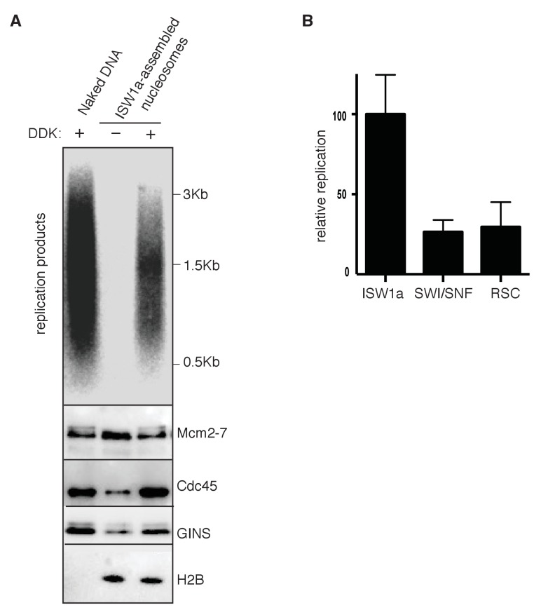
(A) Comparison of naked and nucleosomal DNA templates in the reconstituted replication assay. Comparison of replication of naked DNA and ISW1a templates with or without DDK using a fully reconstituted replication assay. Replication products (top panel) and Mcm2-7, Cdc45, GINS and H2B template association (lower panels) were assayed as in Figure 4D. (B) Comparison of nucleosomal DNA assembled with different CRE in the reconstituted replication assay. Quantification of the ISW1a, SWI/SNF and RSC templates replication products in the presence of DDK using reconstituted replication assay. Reconstituted replication assays performed as described in Figure 4D. Quantified as in Figure 3B and the material and method section. Error bars indicate standard deviation of three biological replicates (n = 3).
Figure 4—figure supplement 1—source data 1. Raw values used in the quantification of Figure 4B (n = 3).
DOI: 10.7554/eLife.22512.018
Figure 4—figure supplement 2. Nucleosomal template assembled without CRE are able to replicate.

Figure 4—figure supplement 2.

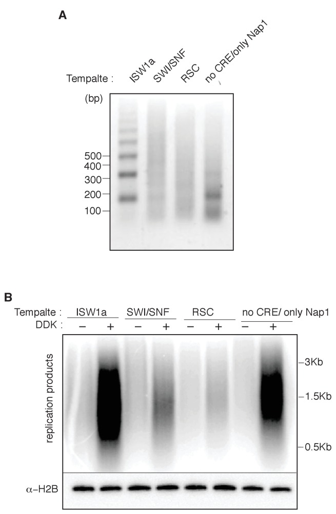
(A) Nucleosomes were assembled with either ISW1a, SWI/SNF, RSC. or no CRE, with bead-coupled ARS1-containing linear DNA, yeast histone octamers and Nap1. Nucleosome assembly was assessed after partial MNase digestion as in Figure 2A. (B) Comparison of ISW1a, SWI/SNF, RSC or no CRE nucleosomal DNA templates in the reconstituted replication assay with or without DDK. H2B template association (lower panels) were assayed as in Figure 4D.