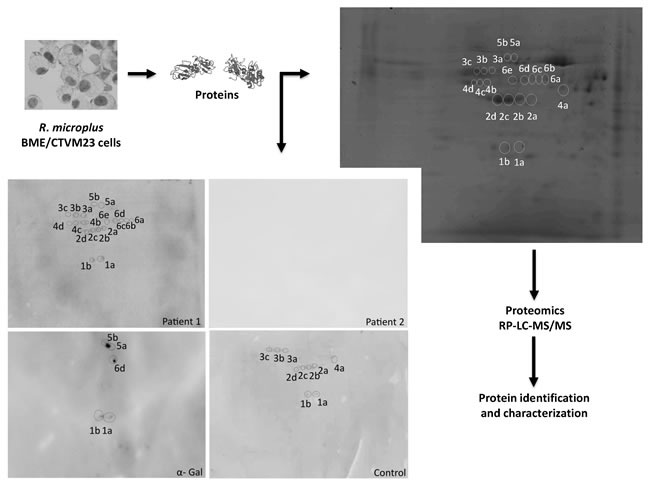
Figure 4.

Rhipicephalus tick proteins recognized by IgE in patient 1 and control sera and by anti-α-Gal IgE antibodies. The R. microplus BME/CTVM23 tick cell proteins were extracted and analyzed by 2-D Western blot using patients and control sera and anti-α-Gal antibodies. The protein spots of interest recognized by patients or control sera and by anti-α-Gal antibodies were manually excised from the stained gel and used for proteomics analysis. The same settings were used for all four panels in which proteins were resolved by isoelectrical focusing at pH 3-11 followed by 12% SDS gel electrophoresis in the second dimension with 140-15 kDa molecular weight range.
