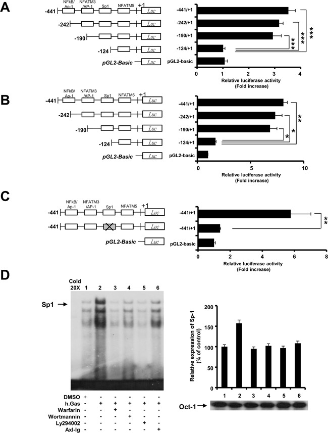
Figure 3. Transcriptional activation of Sp1 regulates Axl-mediated LIGHT expression.

(A and B) Jurkat and 293T cells were co-transfected with plasmids as described in the Materials and Methods. Luciferase activity was measured in Jurkat and 293T cells stimulated with 1μg/ml of rhGas6 for 24h. (C) Site-directed mutagenesis was performed on Sp1 binding site of LIGHT promoter followed by the luciferase assay in 293T cells stimulated with rhGas6. Data are represented as the mean ± SEM from three independent experiments (*, P < 0.05; **, P < 0.01, ***, P < 0.001). (D) Jurkat-Axl cells were pretreated with signal blockers as described in Figure 2 followed by assessment of the DNA-binding activity of Sp1 by EMSA after stimulation with 1μg/ml of rhGas6 for 3h. Oct-1 was used as an internal protein loading control. Cold 20x represents a 20-fold excess of unlabeled Sp1 probe for competition analysis.
