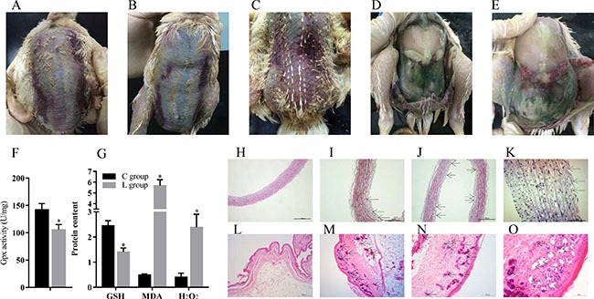
Figure 1. Se deficiency induce exudative diathesis and HE staining for broiler chick veins and skins.

(A–C) The typical exudative diathesis; (D, E) Necropsy photograph of Se deficient broilers; (F) Effect of Se deficiency on GPx; (G) Effect of Se deficiency on GSH, MDA and H2O2. The unit of GPx, GSH, MDA and H2O2 were U mg−1, mg prot−1, nmol·mgprot−1 and mmol·gprot−1; (H) The histopathological analysis in vein of control group; (I–K). The histopathological analysis in vein of Se deficiency group, the arrows in black point to the location of the lesion; (L) The histopathological analysis in skin of control group; (M–O) The histopathological analysis in skin of Se deficiency group, the arrows in black point to the location of the lesion. Each value represented the mean ± S.D. of three individuals. *P < 0.05 versus control group.
