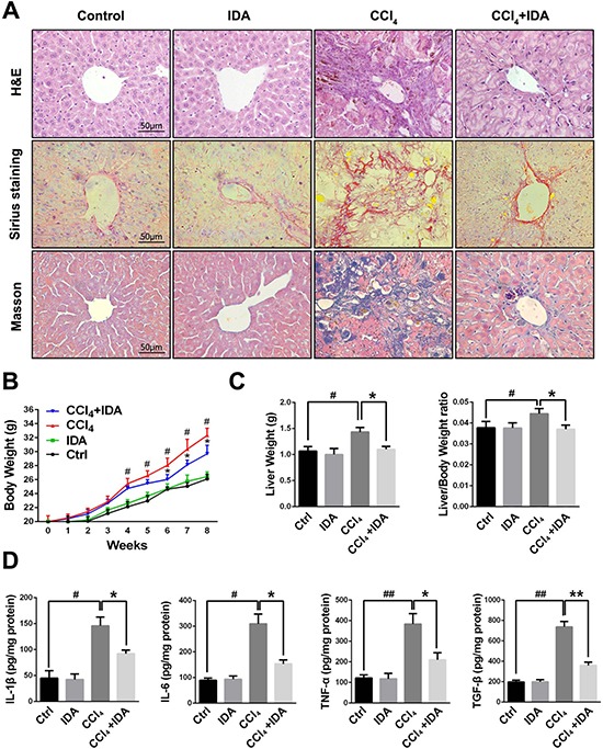
Figure 1. IDA attenuates the progression of liver fibrosis in CCl4-treated mice.

(A) Liver fibrosis as assessed by hematoxylin and erosin (H&E), Sirius red Staining and Masson trichrome staining (n = 6 in each group). Fibrosis is represented by collagen deposition (red color area) in Sirius Staining and (blue color area) in Masson's trichrome staining. (B) Trends in the body weights of C57BL/6 mice were monitored at 1 week intervals throughout the 8 weeks of CCl4 treatment. (C) The liver weights and liver/body weight ratio at 8 weeks were measured. (D) The protein levels of IL-1β, IL-6, TNF-α and TGF-β in liver tissue homogenates were measured by ELISA. The experiments were repeated for three times and data are represented as mean ± SEM. #p < 0.05 versus Control; ##p < 0.01 versus Control; *p < 0.05 versus CCl4; **p < 0.01 versus CCl4.
