Figure 2. Micrograph of transfected cells.

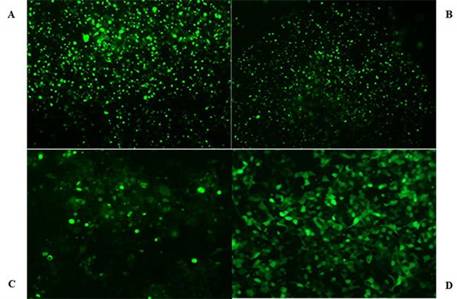
HEK293 transfected cells overexpressing OCT2 (A), Mrp2 (B), Pgp (C), and HEK293-vector cells (D) were photographed by fluorescent microscope.

HEK293 transfected cells overexpressing OCT2 (A), Mrp2 (B), Pgp (C), and HEK293-vector cells (D) were photographed by fluorescent microscope.