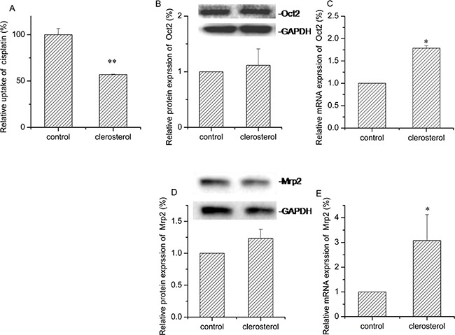
Figure 4. Effect of clerosterol on Mrp2 and Oct2 in BRL cells.

(A) Cisplatin uptake (DDP) was analyzed by HPLC after the BRL cells were incubated with clerosterol for 24 h at 37°C, and then incubated with 50 μg/mL of cisplatin for another 4 h at 37°C. Oct2 protein expression (B) and Mrp2 protein expression (D) in the BRL cells treated with clerosterol for the indicated period of time, determined by Western blot analysis. GAPDH was used as a loading control. Oct2 mRNA expression (C) and Mrp2 mRNA expression (E) was measured by RT-qPCR analysis. Error bars indicate SD. *p < 0.05, **p < 0.01 compared with the control.
