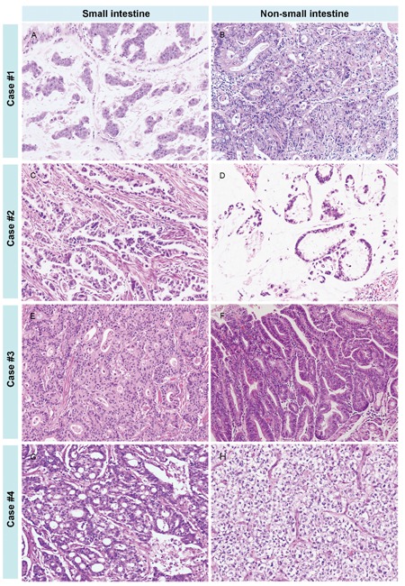
Figure 3. The various histologic features of SIACs and matched metachronous tumors.

In case number 1, the SIAC A. was mucinous, while the metachronous colorectal cancer B. was tubular without a mucin component. In case number 2, the SIAC C. was tubular, but the metachronous colorectal tumor D. was mucinous. In case number 3, the SIAC E. was moderately differentiated and tubular, while the metachronous early gastric cancer F. was a well-differentiated tubular adenocarcinoma. In case number 4, the SIAC G. was moderately differentiated and tubular, and the metachronous brain tumor H. was an anaplastic oligodendroglioma. (All images, 200× magnification.)
