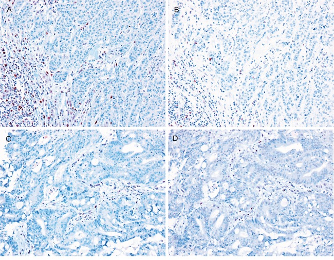
Figure 5. Representative images of MMR protein expression in LS-related SIACs.

Loss of A. MLH1, B. PMS2, C. MSH2, and D. MSH6 protein expression was observed in LS-related SIACs. Lymphocytes are used as internal positive controls. (All images, 200× magnification.)
