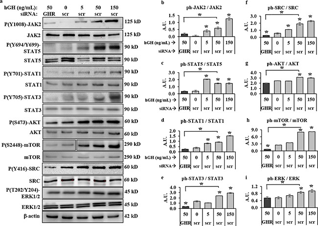
Figure 3. GH-excess promotes and GHRKD attenuates multiple critical intracellular signaling pathways in human melanoma cells.

a. Representative images of western blot (WB) analyses of phosphorylation levels b. JAK2, c. STAT5, d. STAT1, e. STAT3, f. SRC, g. AKT, h. mTOR and i. ERK1/2, in excess human-GH treated or GHRKD human melanoma cell lysates. SK-MEL-28 cells, 24 hr post-transfection with either scramble (scr)-siRNA or GHR-siRNA were treated for ten mins with GH and lysed as described. WB was performed using appropriate antibodies. Densitometry analyses of individual blots was performed using ImageJ software and the ratio of phosphorylated vs. total protein levels against untreated scr-siRNA transfected controls. Overall, excess GH increased while GHRKD decreased phosphorylation states. Similar results for MALME-3M, MDA-MB-435 and SK-MEL-5 human melanoma cells are presented in Supplementary Figure 3. Blots from individual experiments were quantified and the mean of three blots per antibody was taken. Protein levels were normalized against expression of β-actin. [*, p < 0.05, Students t test, n = 3].
