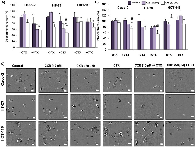
Figure 8. Combined Celecoxib/Cetuximab treatment in colon cancer cells impairs colonosphere formation capability.

Colon cancer cells were pre-treated with 10 μM or 50 μM Celecoxib (CXB) as a single agent or in combination with 100 μg/ml Cetuximab (CTX) for 48h, and then cells were seeded at clonal density with serum free medium in low-adherence plates. After seven days, A. the number, B. size and, C. appearance of formed colonospheres were evaluated by light microscopy. Spheres number and size were quantified with the incuCyte ZOOM® Software 2015A. (Final magnification: X40, scale bar corresponds to 100 microns). Data are means ± SEM of three independent experiments (*p <0.05, compared with the control; # p<0.05, compared with cetuximab-treated cells).
