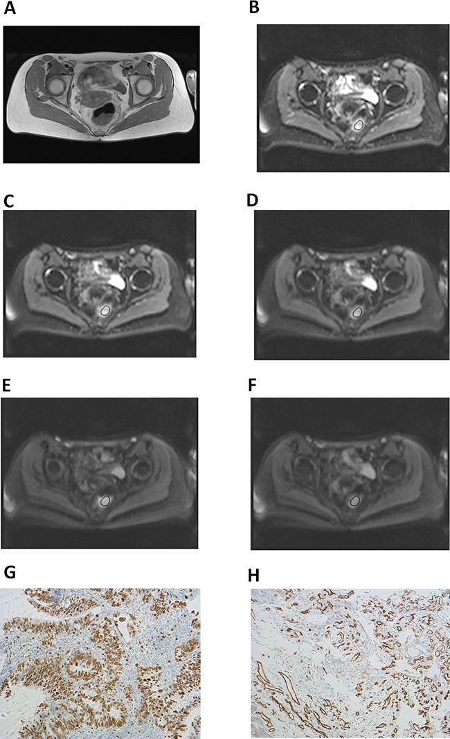
Figure 4. Imaging and histopathological findings in rectal cancer.

(A) T2w image showing a large rectal mass. Histopathological investigation after endoscopic biopsy confirmed the diagnosis of a moderately differentiated adenocarcinoma. Tumor stage: T3 N1 M0. (B-F) DW imaging findings: b0 (B), b50 (C), b200 (D), b500 (E), and b1000 (F). Estimated IVIM parameters are as follows: ADC=1. 02 × 10−3 mm2s−1, D=0.71 × 10⁻³ mm2s−1, f=28%, D*=16.60 × 10⁻³ mm2s−1, and fD*=5.03. (G) Immunohistochemical stain (MIB-1 monoclonal antibody). Histopathological parameters are as follows: Ki 67 index=75%, cell count=1390, total nucleic area= 225375.10 μm2, and average nucleic area=162.23 μm2. (H) Immunohistochemical stain (CD 31monoclonal antibody). Estimated microvessel parameters are as follows: stained vessel area=70555.98 μm2, total vessel area=84787.53 μm2, mean vessel diameter=11.97 μm, and vessel count=210.
