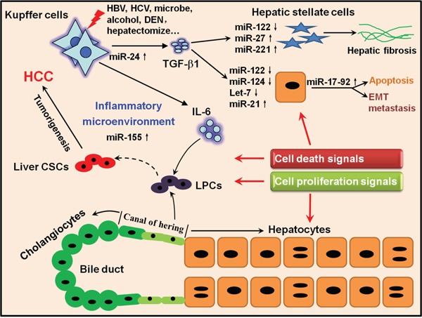
Figure 6. A hypothetical illustration delineating the connection between activation of inflammatory pathways, miRNAs and liver tumorigenesis.

Once extrinsic stimuli such as HBV/HCV, alcohol and DEN damage the liver, Kupffer cells can be activated and produce several inflammatory cytokines such as IL-6 and TGF-β1. On one hand, IL-6 can stimulate LPCs residing in the canal of hering to proliferate to restore the injured liver; however, if gene mutations happen to proliferating LPCs, they will have the potential to develop to CSCs. On the other hand, TGF-β1 can act on HSCs and activated HSCs proliferate and generate ECM to reconstitute the liver and promote hepatic fibrosis if the dynamic balance of ECM synthesis and decomposition is disrupted. Meanwhile, TGF-β1 can also stimulate hepatocytes to respond to either cell death or proliferation signals under different conditions. Several miRNAs such as miR-122, miR-155 and miR-21 could join to regulate correlated pathologic processes. All the cytokines, miRNAs and other inflammatory mediators together generate an inflammatory microenvironment which will amplify the oncogenic mutations and self-reinforce the pro-inflammatory signals, finally leading to the irreversible liver tumorigenesis.
