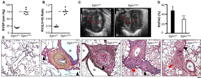
Figure 1. Spontaneous severe PAH with obliterative pulmonary vascular remodeling in Egln1Tie2 mice.

(A) Marked increase of RVSP. (B) Unprecedented RV hypertrophy. (C) Echocardiography demonstrating marked thickness of RV wall (double arrow) and enlarged RV chamber. LV, left ventricle. (D) Decreased RV fraction area indicating RV dysfunction. (E) Representative micrographs of Russel-Movat pentachrome staining demonstrating thickening of the intima, media, and adventitia; occlusion of the large and small vessels (black arrowheads); and plexiform-like lesions (red arrowheads) in 3.5-month-old Egln1Tie2 mice. Data are expressed as mean ± SD. ***, P < 0.001. (Adapted from ref. 8).
