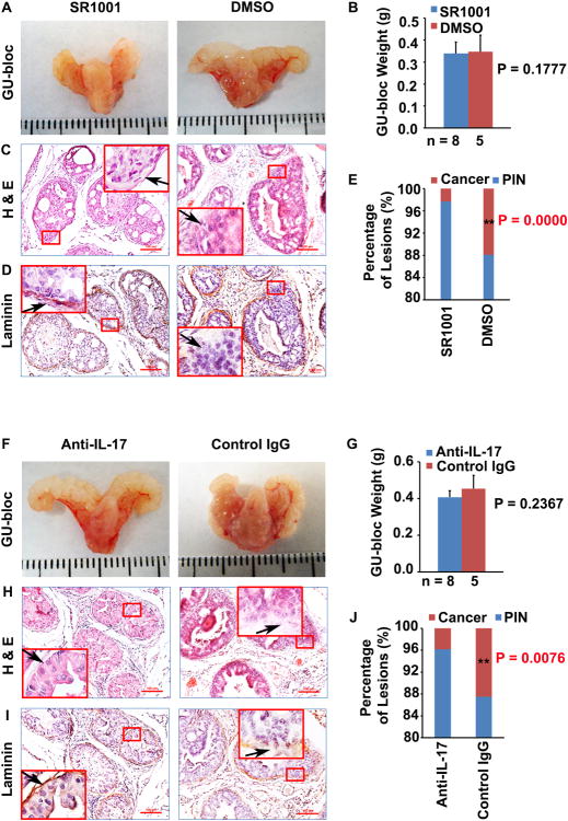
Fig. 1. SR1001 or IL-17 antibody decreases the formation of invasive prostate adenocarcinoma in mice.
A: Representative photographs of the GU-blocs in SR1001 and DMSO (control)-treated mice. B: GU-bloc weight; the number of animals in each group is shown under the abscissa. C and D: Representative sections of dorsolateral prostatic lobes stained with H&E and anti-laminin; original magnification, ×100 for photomicrographs and ×400 for inserts; arrow indicates a micro-invasive site where continuity of laminin staining is broken in control mice and a non-invasive site where the continuity of staining is not broken in SR1001-treated mice; the tissue sections in D were consecutive sections to C. E: Percentages of PIN and cancer (i.e., micro-invasive prostate adenocarcinoma) in ventral, dorsal and lateral prostatic lobes of SR1001-treated and control mice; n = 8 and 5 animals in SR1001 and DMSO treatment groups respectively, **P < 0.01 compared with control mice. F: Representative photographs of the GU-blocs in IL-17 antibody and control IgG-treated mice. G: GU-bloc weight; the number of animals in each group is shown under the abscissa. H and I: Representative sections of dorsolateral prostatic lobes stained with H&E and anti-laminin; original magnification, ×100 for photomicrographs and ×400 for inserts; arrow indicates a micro-invasive site where continuity of laminin staining is broken in control IgG-treated mice and a non-invasive site where the continuity of staining is not broken in IL-17 antibody-treated mice; the tissue sections in I were consecutive sections to H. J: Percentages of PIN and micro-invasive cancer in ventral, dorsal and lateral prostatic lobes of IL-17 antibody and control IgG-treated mice; n = 8 and 5 animals in IL-17 antibody and control IgG treatment groups respectively, **P < 0.01 compared with control mice.

