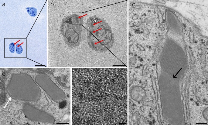
Fig. 1.
Ultrastructure of intramitochondrial crystals in articular chondrocytes of juvenile horses. a Semithin resin section stained with toluidine blue showing three chondrocytes containing two types of intensively stained structures inside the cytoplasm: spherical lipid droplets and crystals (arrows). The square indicates the area of b. b TEM image showing the same double chondron as in a bearing electron dense lipid droplets (L) and glycogen (G) and protein crystals (arrows). The latter have elongated and homogenous structures and a slightly darker appearance than the cytoplasm. The cell on the right contains several crystal transections. The square indicates the area of magnification of image c. c Crystal consisting of three compartments. A striated substructure is visible in the middle compartment. A less dense striated zone forms the transition to the lower compartment (arrow). The whole crystal is surrounded by a double membrane. d Three crystals with different geometric shapes surrounded by a double membrane. In the outer left-hand crystal, cristae are visible between the crystal and the mitochondrial membrane (arrow). e High magnification of the crystal structure showing the individual protein units. Scale bars are a 20 µm, b 4 µm, c 250 nm, d 500 nm and e 2 nm

