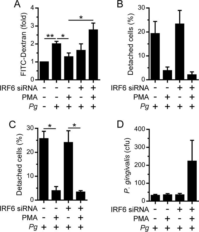
FIG 6.
Regulation of oral keratinocyte barrier function by IRF6. OKF6 cells were transfected with an IRF6 (+) or control (–) siRNA. Thereafter, the cells were treated with 100 ng/ml PMA (+) or 0.1% DMSO (–) for 24 h. (A) Cells growing on a Transwell insert were incubated with P. gingivalis (MOI of 100:1) and 1 mg/ml FITC-dextran for 60 min, and FITC-dextran levels in the lower chamber were then measured (n = 4). (B and C) Cells growing in a 12-well plate were cultured with P. gingivalis (MOI of 100:1) for 24 h. (B) Detached cells were collected and enumerated (n = 2). (C) The remaining adherent cells in the experiment performed as described for panel B were incubated in PBS-EDTA for 60 min, and the numbers of detached cells were then enumerated (n = 2). In panels B and C, the numbers of detached cells are presented as a percentage of the total (detached plus attached) cell number. (D) Cells growing in a 12-well plate were cultured with P. gingivalis (MOI of 100:1) for 2 h, and bacterial invasion was then measured by antibiotic protection assay (n = 4). **, P < 0.01; *, P < 0.05.

