
FIG 5.
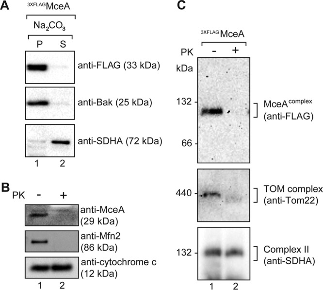
MceA is localized to the mitochondrial outer membrane. (A) Mitochondria isolated from cells infected with Coxiella expressing 3×FLAGMceA were subjected to alkaline extraction using 100 mM Na2CO3 (pH 11). The membrane-integrated protein fraction (the pellet fraction [P]) and the peripheral membrane protein fraction (the supernatant fraction [S]) were obtained by ultracentrifugation and subsequently analyzed by SDS-PAGE and immunoblotting using the indicated antibodies. (B and C) Mitochondria isolated from cells infected with Coxiella expressing 3×FLAGMceA were either left untreated (lanes 1) or treated with PK (50 μg/ml) (lanes 2) and either analyzed by SDS-PAGE (B) or solubilized in digitonin-containing buffer and analyzed by BN-PAGE (C). Immunoblotting was performed with the indicated antibodies.