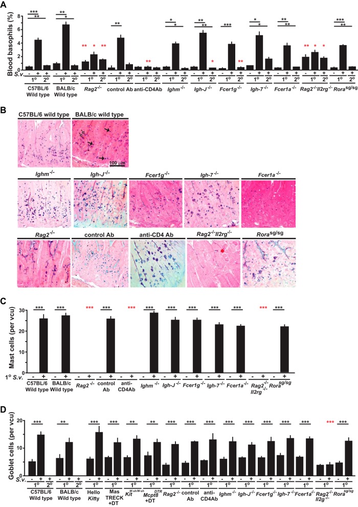
FIG 4.
Changes in numbers of basophils, mast cells, and epithelial goblet cells in mice deficient in various components of immune responses during primary and secondary S. venezuelensis infections. (A) Blood was collected from each mouse during all of the experiments performed as described for Fig. 3A to J before infection or 10 to 12 days after 1o or 2o S. venezuelensis infections and stained with anti-IgE and anti-CD49b. Percentages of basophils in live cells are shown as bar graphs with means + SEM. ***, P < 0.0005; **, P < 0.005; *, P > 0.05; no asterisks, P > 0.05. Black asterisks indicate the statistical significance of results of comparisons between values for uninfected mice and 1o or 2o infected mice in the same group (as indicated by bars). Red asterisks indicate the statistical significance of results of comparisons between each type of mutant or anti-CD4 Ab-treated mouse and the corresponding control mice for that condition. (B to D) Jejunums were collected 14 days after the start of 1o infections, and sections of jejunum were stained as described for Fig. 2B (in the panel showing the histology in BALB/c wild-type mice, some of the mast cells are indicated with filled arrows and some of the goblet cells are indicated with open arrows) (B), and numbers of mast cells (C) or goblet cells (D) per villus crypt unit (vcu) are shown as bar graphs (n = 4 to 12 each; data were combined from 2 or 3 experiments). Wild-type values for comparisons to those from mutant mice were combined from corresponding controls of the same strain (C57BL/6 or BALB/c).

