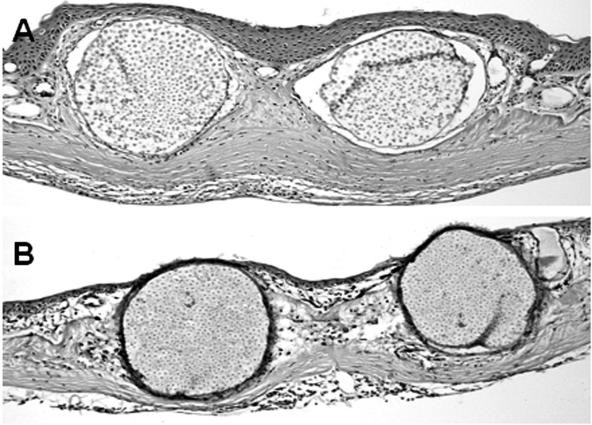
FIG. 1.
Hematoxylin- and eosin-stained micrographs of the skin infected by Amphibiocystidium ranae in Rana esculenta (A) and Rana lessonae (B). The intradermal cysts were located subjacent to the epidermis, which is more hyperplasic in R. esculenta than in R. lessonae. The A. ranae endospores appear more developed in the skin sample of R. esculenta than in R. lessonae. A, ×15; B, ×10.

