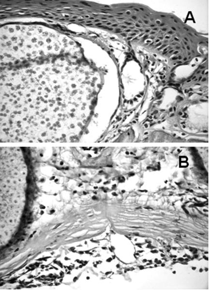
FIG. 2.
Detail of cysts and endospores of Amphibiocystidium ranae in infected Rana esculenta (A) and Rana lessonae (B). Inflammatory cells, such as eosinophils and neutrophils, are also observed near the cysts. Hematoxylin and eosin, ×40. Note that the endospores within the cysts of A. ranae in R. esculenta (A) appear more developed than in R. lessonae (B).

