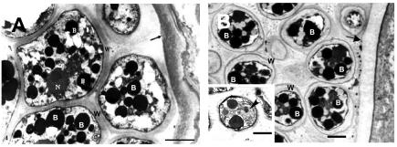
FIG. 3.
(A) Transmission electron micrograph of Amphibiocystidium ranae mature sporangium (cyst) containing endospores from an infected frog. The arrow points to the sporangium cell wall; B depicts multiple electron dense bodies; N, nucleus; W, capsule of the endospores. Bar, 2 μm. (B) Rhinosporidium seeberi from a Sri Lankan man with rhinosporidiosis. The arrow points to the mature sporangium cell wall. B, electron-dense bodies; W, endospore wall. Bar, 2 μm. Note the striking ultrastructural similarities between these two pathogenic mesomycetozoeans. The left lower insert in panel B shows a transmission electron micrograph of R. seeberi endospores with a prominent nucleus and a nucleolus (arrowhead). Bar, 2.5 μm.

