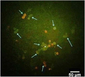
Fig. 3.

FITC-BCG binding and internalization by A549 cells. A549 cells were exposed to FITC-BCG for 20 min in standard culture conditions. After washing, the green surface fluorescence of bound bacteria was quenched for 3 min incubation on ice with Trypan blue and cytospun cells observed by fluorescence microscopy. The arrows point to the quenched cell-bound bacteria (red hollow circle) and internalized particles (green), which remain green, as they were not exposed to Trypan blue. The image was captured with a 40× objective and representing photo selected from 3 independent experiments
