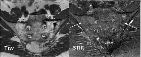
Fig. 2.

MRI of bony pelvis. The MRI of the patient’s pelvis reveals bony fusion of the bilateral sacroiliac joints with thickening of the cortex of the left iliac bone and bony trabeculation of the sacrum and left iliac bone. Degenerative changes are seen in the visualized lower lumbar spine. There is a hyperintense signal intensity in the left sacral ala which is likely due to micro trabecular insufficiency fractures
