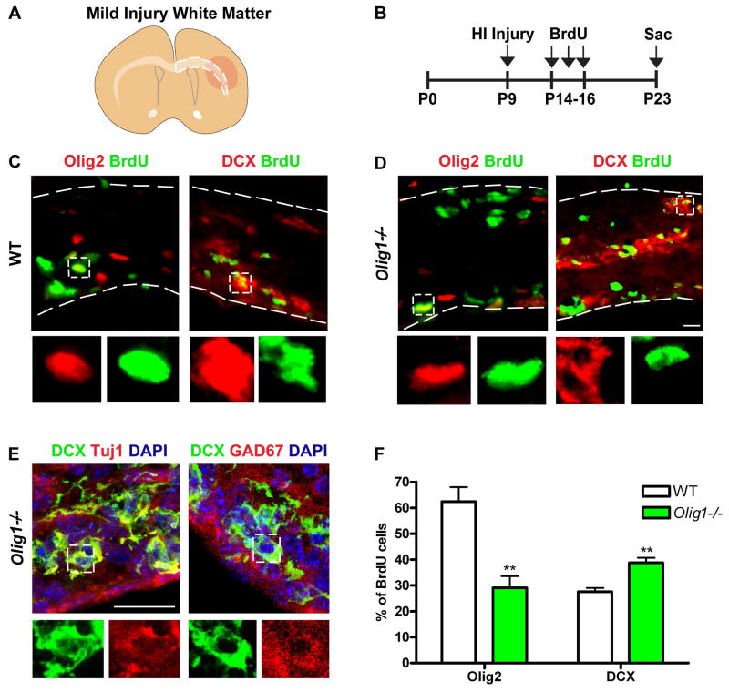
Figure 2. Ectopic production of interneurons and depressed proliferation of OPCs in white matter of Olig1-null animals post injury.
(A) Cartoon illustrating subcortical white matter in mild neonatal HI injury. (B) Experimental timeline for BrdU administration following neonatal HI injury. (C–D) Representative image of DCX (red) or Olig2 (red) and BrdU (green) immunostaining in the subcortical white matter of a wild type and Olig1-null animal two weeks post injury. Note the increased number of proliferating DCX-positive cells in the subcortical white matter of an Olig1-null animal. (E) Confocal projections showing that DCX (green) colocalizes with cellular Tuj1 (red) and GAD67 (red) in the subcortical white matter of Olig1-null animals. (F) Quantification of the percentage of BrdU cells expressing DCX or Olig2 in the subcortical white matter of wild type and Olig1-null animals. Data are mean ± s.e.m; n=3 animals per genotype; **P<0.01; Student’s two-tailed t test. (D,E) scale bar = 20μm.

