
Figure 3. Pab1 demixing proceeds via liquid-liquid phase separation and gel formation, modulated but not caused by its low-complexity region.
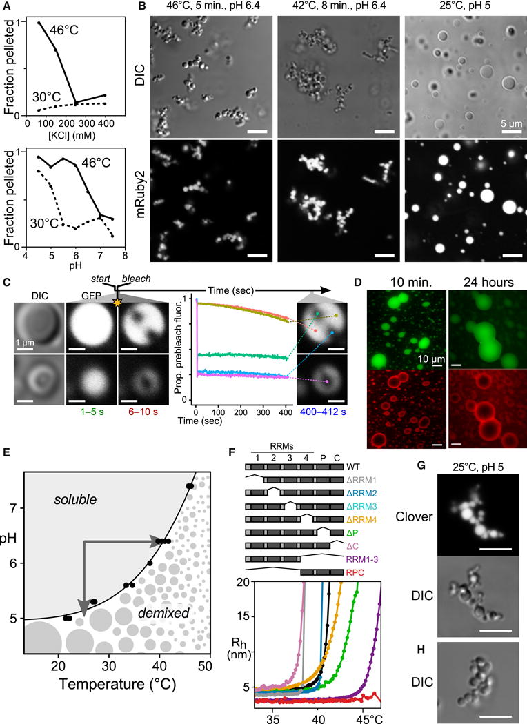
A, Demixing of purified Pab1 is sensitive to ionic strength and pH (Fig. S3) B, Morphology of 15 μM Pab1-mRuby2 assemblies. C, Fluorescence recovery after photobleaching of Pab1 droplets. D, Sequentially assembled two-color droplets remain unmixed after 24 hours. E, The Tdemix of Pab1 measured at different pH values defines a phase boundary. F, Pab1 domain deletions and corresponding DLS temperature ramps (Table S1). G, H, Morphology of Pab1ΔP quinary assemblies. G, 15 μM 100:3 Pab1ΔP:Pab1-Clover; H, 15 μM Pab1ΔP alone.