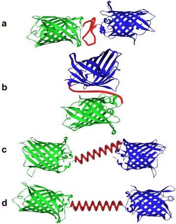
Fig. 28.

Schematic illustrations of various conformations of the fusion proteins. a EBFP (blue) and EGFP (green) are situated in a straight line, with the flexible linker (red) between the two domains. b EBFP and EGFP reside side by side, for the most compact conformation with the flexible linker. c The helical linker connects EBFP and EGFP diagonally. d The helical linker and the long axes of EBFP and EGFP are situated in a straight line
(Figure adapted with permission from: Ref. [347]. Copyright (2004) John Wiley & Sons)
