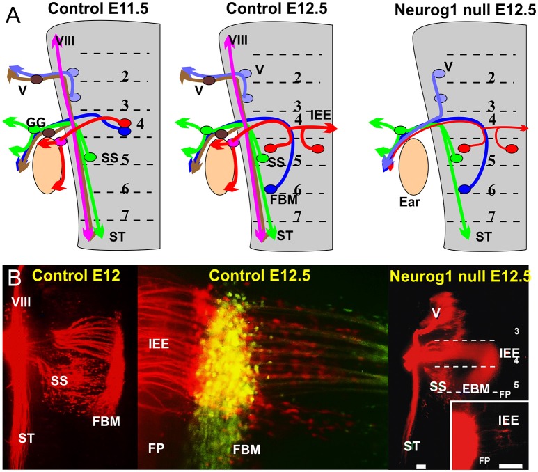
Figure 2.
(A) The distribution of afferents and efferents, including the FBM and IEE is shown for a flat mounted hindbrain of an 11.5 day old mouse embryos prior to migration and a 12.5 day mouse embryo after migration. Note that the trigeminal afferents (V) and efferents enter/exit at r2, superior salvatory (parasympathetic visceral efferents) are in r5 and exit with visceral afferents running in the solitary tract (ST). FBM and IEEs in r4 and ran with afferents of the geniculate ganglion (GG) and inner ear afferents (VIII). Absence of Neurog1 eliminates all neural crest derived sensory neurons but retains the visceral sensory neurons of the GG to project to the solitary tract. Trigeminal efferents cannot exit through r2 but instead exit through r4 together with FBM and reduced numbers of IEEs. (B) The photomicrographs show the distribution of FBMs, SS and IEEs at E12 and the appearance of bilateral IEE overlapping with FBM at E12.5 (center). In Neurog1 mutant mice facial labeling also labels trigeminal motor neurons as well as FBM, SS and a reduced number of bilateral IEEs. Only the GG afferents forming the solitary tract remain. Bar indicates 100 um. Modified after Ma et al. (2000) and Fritzsch and Nichols (1993).

