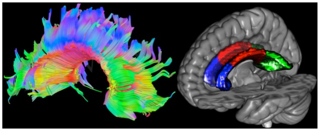
Figure 2.

Left: tracts of the corpus callosum (CC) projected on the diffusion weighted image of one healthy control (HC) participant. Red, green and blue represent left to right, anterior to posterior and superior to inferior directions, respectively. Right: masks of the CC projected on the Collins MNI-reference brain. The genu is colored in blue, the body in red and the splenius in green.
