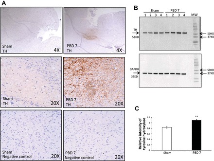
Fig. 5.

A: sympathetic activity is increased in brain after burn injury. Immunohistochemical detection of tyrosine hydroxylase (TH) in mouse brain after PBD7 is compared with sham. TH is represented as a dark brown reaction product of DAB oxidation in the presence of peroxidase and hydrogen peroxide at the site of enzymatic activity. All slides were counterstained with hematoxylin. Images were captured by light microscopy at ×4 and ×20 magnification using EVOS FL Cell Imaging System. Experiment was repeated 3 times with n = 4 mice per group. B: Western blot analysis of TH bands at 58 kDa and GAPDH bands at 37 kDa in brain of PBD7 mice compared with sham. MW, molecular weight. C: TH protein levels based on densitometry expressed as means ± SE relative to GAPDH as a loading control. Bar graph represents means ± SE, containing 7 mice/group showing increase in TH protein expression in the brains of PBD7 mice. **P < 0.01 vs. sham by one-way ANOVA.
