Fig. 6.
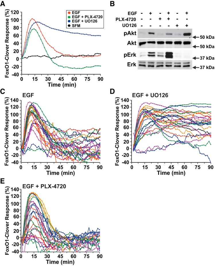
ERK activity negatively regulates Akt signaling by EGF. A: time course of the relative translocation response of the FoxO1-clover reporter in C3H10T1/2 cells incubated in SFM and then exposed to SFM or EGF (2.1 nM) ± different inhibitors for 90 min. Population averages are presented (n = 50 cells/incubation). The relative responsiveness of the reporter protein in each cell was normalized to the value at the start of imaging during incubation in SFM and scaled to the average peak EGF response. B: expression of pAkt, total Akt, pERK, and total ERK by immunoblotting using whole cell protein lysates from C3H10T1/2 cells after exposure to SFM or EGF plus the indicated inhibitors for 15 min. Molecular mass markers are indicated to the right of each immunoblot. C: time course results for each of 25 individual cells incubated with EGF. D: time course results for each of 25 individual cells incubated with EGF plus U-0126 (10 μM). E: time course results for each of 25 individual cells incubated with EGF and PLX-4720 (10 μM). Cells were imaged every 2 min during each treatment period. The nuclear intensity of the reporter in each cell was normalized to its value at the start of imaging during incubation in SFM and scaled to the average peak EGF response.

