Fig. 7.
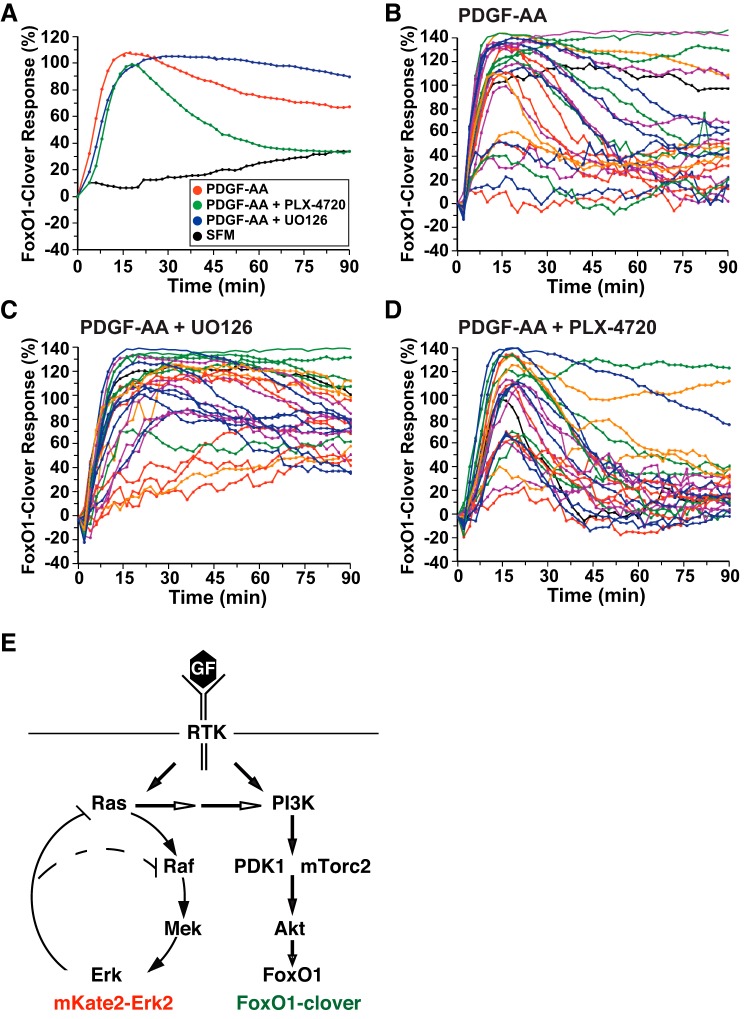
ERK activity negatively regulates Akt signaling by platelet-derived growth factor (PDGF)-AA. A: time course of the relative translocation response of the FoxO1-clover reporter in C3H10T1/2 cells incubated in SFM and then exposed to SFM or PDGF-AA (1.4 nM) ± different inhibitors for 90 min. Population averages are presented (n = 50 cells/incubation). The relative responsiveness of the reporter protein in each cell was normalized to the value at the start of imaging during incubation in SFM and scaled to the average peak response. B: time course results for each of 25 individual cells incubated with PDGF-AA. C: time course results for each of 25 individual cells incubated with PDGF-AA plus U-0126 (10 μM). D: time course results for each of 25 individual cells incubated with PDGF-AA and PLX-4720 (10 μM). Cells were imaged every 2 min during each treatment period. The nuclear intensity of the reporter in each cell was normalized to its value at the start of imaging during incubation in SFM and scaled to the average peak response. E: schematic of interrelationships between the Ras–Raf–Mek–ERK and phosphatidylinositol (PI) 3-kinase–Akt signaling pathways. Feedback inhibition by ERK activity on Raf and Ras and activation of PI 3-kinase by Ras are indicated. The locations of mKate2-ERK2 and FoxO1-clover as sensors for these pathways are shown.

