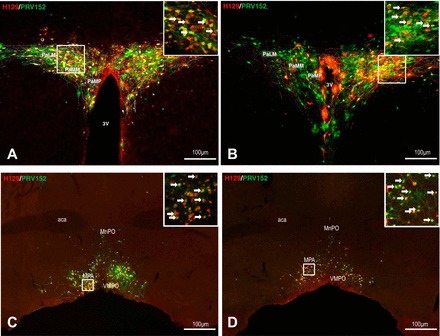
Fig. 6.

Left: low and high (inset) magnification of the photomicrographs illustrating single H129 (red), single PRV152 (green), and colocalized H129 + PRV152 immunolabeling (yellow) cells in the PVH (A) and MPA (C) in the forebrain following H129 microinjections intra-IBAT and PRV152 intra-IWAT. Right: equitable immunolabeling in the PVH (B) and MPA (D) after the viruses were switched. Arrows indicate double-labeled neurons. Scale bar = 100 µm. 3V, third ventricle; aca, anterior commissure; MnPO, median preoptic nucleus; MPA, medial preoptic area; PaLM, paraventricular hypothalamic nucleus, lateral magnocellular part; PaMM, paraventricular hypothalamic nucleus, medial magnocellular part; PaMP, paraventricular hypothalamic nucleus, medial parvicellular part; VMPO, ventromedial preoptic nucleus.
