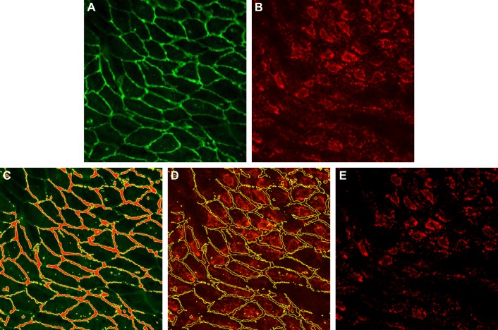
Fig. 1.
Measurement of background intensity. A: VE-cadherin channel. B: original cyclooxygenase-2 (COX-2) channel. C: using the threshold function in ImageJ, the junction area is selected in the VE-cadherin channel. In the junction area, COX-2 is expected to be low or absent. D: the selection is then transferred to the COX-2 channel and the mean intensity within the selection is taken as the background intensity. E: background-corrected COX-2 channel.

