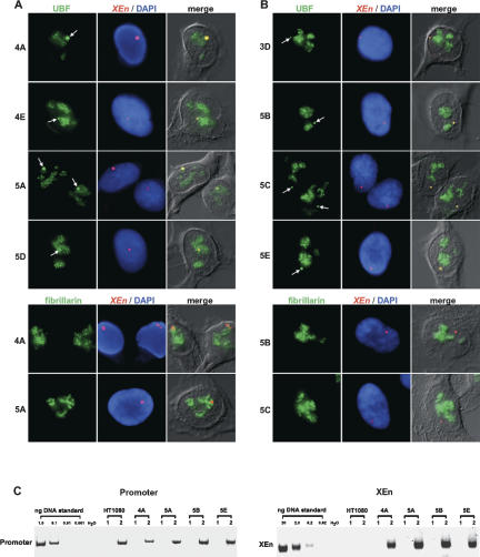
Figure 2.
UBF binds to XEn arrays in vivo. (A) Combined immuno-FISH on clones 4A, 4E, 5A, and 5D containing XEn arrays on the short arms of acrocentric chromosomes. (Top four rows) UBF was visualized using FITC-conjugated affinity-purified antibodies. (Bottom two rows) Fibrillarin was visualized in clones 4A and 5A using monoclonal antibody 72B9 combined with Cy2-labeled α-mouse secondary antibodies. XEn arrays were visualized with a spectrum red-labeled probe. For each clone, the row shows antibody staining (left), the XEn probe with DAPI counterstain (center), and the merge of antibody and XEn signals with a DIC image of the cell (right). Arrows indicate XEn arrays. (B) Combined immuno-FISH on clones 3D, 5B, 5C, and 5E containing XEn arrays on submetacentric chromosomes. Performed and illustrated as above. (C) Chromatin immunoprecipitation demonstrates that UBF is bound to XEn arrays. ChIP was performed on soluble chromatin fractions prepared from HT1080 parental cells and clones 4A, 5A, 5B, and 5E using control IgG (lanes labeled 1) or affinity-purified α-UBF antibodies (lanes labeled 2). PCR reactions were performed using primers specific for the human ribosomal gene promoter (left) or the XEn array (right). Serial dilutions of a cosmid comprising the entire human ribosomal repeat or genomic DNA isolated from clone 4A were used as standard templates in PCRs with promoter and XEn primers respectively.

