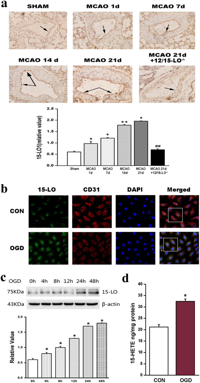
Figure 1. 15-lipoxygenase (15-LO) expression is increased in brain artery endothelium of post-stroke mice and OGD-induced BMVECs.
(a) Immunohistochemical staining of 15-LO in brain arteries from WT mice suffered MCAO for different days and 12/15-LO−/− mice (n = 10, *p < 0.05, **p < 0.01, ##p < 0.01). Values are represented as the mean ± S.E.M. (b) BMVECs were fixed and stained with anti-15-LO (green), anti-CD31 (red) and DAPI to stain nuclei (blue). Merged images show 15-LO colocalizes to CD31 (a marker of ECs). Scale bars are 25 μm. Images shown are representative of at least three independent experiments. (c) Quantification of 15-LO protein levels in BMVECs under OGD for different time points (0, 4, 8, 12, 24, 48 hours; n = 4, *p < 0.05). Values are represented as the mean ± S.E.M. (d) The endogenous level of 15-HETE was measured by15-HETE EIA kit in BMVECs. OGD increases the endogenous15-HETE production compared with normoxic conditions (n = 4, *p < 0.05).

